Home

Aşağı in Jane Austen Aksine in vivo staining organ antagonist Büyütme

MitoView™ Fix 640 - Biotium

Biosensors | Free Full-Text | Selective In Vitro and Ex Vivo Staining of  Brain Neurofibrillary Tangles and Amyloid Plaques by Novel Ethylene  Ethynylene-Based Optical Sensors
Biosensors | Free Full-Text | Selective In Vitro and Ex Vivo Staining of Brain Neurofibrillary Tangles and Amyloid Plaques by Novel Ethylene Ethynylene-Based Optical Sensors

In Vivo Click Chemistry Enables Multiplexed Intravital Microscopy - Ko -  2022 - Advanced Science - Wiley Online Library
In Vivo Click Chemistry Enables Multiplexed Intravital Microscopy - Ko - 2022 - Advanced Science - Wiley Online Library
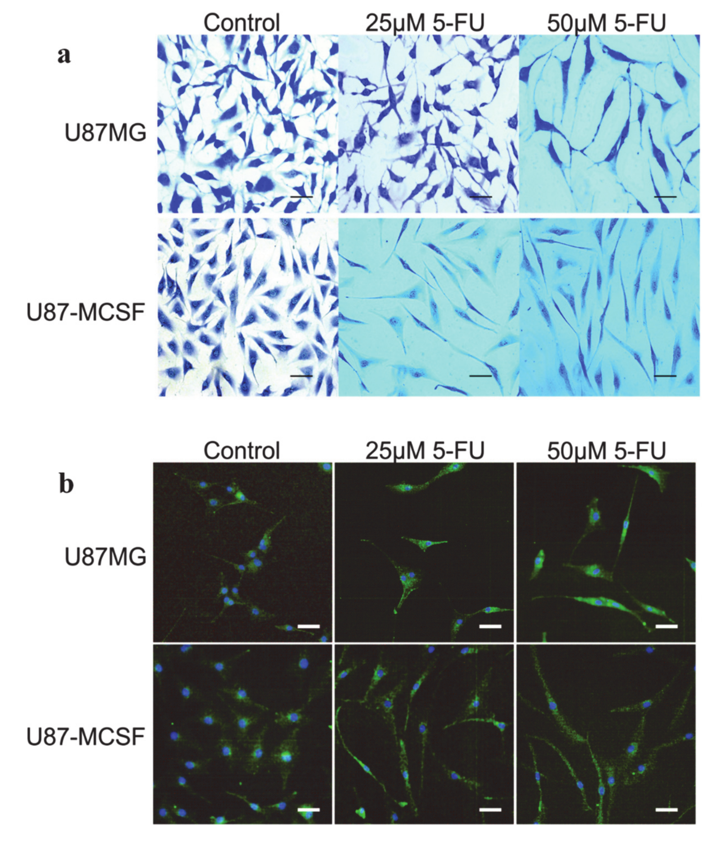
Types of Staining | PDF | Staining | Life Sciences

In vivo staining of the mitochondrion to measure the membrane potential by  Rhodamine 123 dyes.
In vivo staining of the mitochondrion to measure the membrane potential by Rhodamine 123 dyes.

Annexin-vivo | Application Support Knowledgebase | Lab Products & Services  | PerkinElmer
Annexin-vivo | Application Support Knowledgebase | Lab Products & Services | PerkinElmer

Diagnostics | Free Full-Text | Comparative Study Regarding the Properties of  Methylene Blue and Proflavine and Their Optimal Concentrations for In Vitro  and In Vivo Applications
Diagnostics | Free Full-Text | Comparative Study Regarding the Properties of Methylene Blue and Proflavine and Their Optimal Concentrations for In Vitro and In Vivo Applications

JCI Insight - DNA promoter hypermethylation of melanocyte lineage genes  determines melanoma phenotype
JCI Insight - DNA promoter hypermethylation of melanocyte lineage genes determines melanoma phenotype

Immunohistochemistry staining in vivo. (A) In the peri-infarcted area... |  Download Scientific Diagram
Immunohistochemistry staining in vivo. (A) In the peri-infarcted area... | Download Scientific Diagram

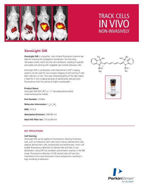
XenoLight DiR - Track Cells In Vivo Non-Invasively - PerkinElmer

In vivo imaging of zebrafish retinal cells using fluorescent coumarin  derivatives | BMC Neuroscience | Full Text
In vivo imaging of zebrafish retinal cells using fluorescent coumarin derivatives | BMC Neuroscience | Full Text

In vivo cardiac findings. (A) H&E staining of longi | Open-i

In vivo two-photon calcium imaging of neuronal networks | PNAS

Double in vivo staining with acridine orange (AO) and ethidium bromide (EB)  as a useful tool for detecting and quantifying the state of dead, dying and  living cells in root meristems of
Double in vivo staining with acridine orange (AO) and ethidium bromide (EB) as a useful tool for detecting and quantifying the state of dead, dying and living cells in root meristems of

Revisiting in vivo staining with alizarin red S - a valuable approach to  analyse zebrafish skeletal mineralization during development and  regeneration – topic of research paper in Biological sciences. Download  scholarly article
Revisiting in vivo staining with alizarin red S - a valuable approach to analyse zebrafish skeletal mineralization during development and regeneration – topic of research paper in Biological sciences. Download scholarly article

H&E staining of in vivo and in vitro skin. The epidermis contains all... |  Download Scientific Diagram
H&E staining of in vivo and in vitro skin. The epidermis contains all... | Download Scientific Diagram

In vivo detection of tau fibrils and amyloid β aggregates with luminescent  conjugated oligothiophenes and multiphoton microscopy | Acta  Neuropathologica Communications | Full Text
In vivo detection of tau fibrils and amyloid β aggregates with luminescent conjugated oligothiophenes and multiphoton microscopy | Acta Neuropathologica Communications | Full Text

In vivo non-invasive staining-free visualization of dermal mast cells in  healthy, allergy and mastocytosis humans using two-photon fluorescence  lifetime imaging | Scientific Reports
In vivo non-invasive staining-free visualization of dermal mast cells in healthy, allergy and mastocytosis humans using two-photon fluorescence lifetime imaging | Scientific Reports

Specific In Vivo Staining of Astrocytes in the Whole Brain after  Intravenous Injection of Sulforhodamine Dyes | PLOS ONE
Specific In Vivo Staining of Astrocytes in the Whole Brain after Intravenous Injection of Sulforhodamine Dyes | PLOS ONE

March 6, 2023 | AI-based staining of biological samples - California  NanoSystems Institute
March 6, 2023 | AI-based staining of biological samples - California NanoSystems Institute

Specific staining of in vivo regenerated tissues at 4 weeks. Samples in...  | Download Scientific Diagram
Specific staining of in vivo regenerated tissues at 4 weeks. Samples in... | Download Scientific Diagram

In vivo propidium iodide (PI) staining assessment in the transplanted... |  Download Scientific Diagram
In vivo propidium iodide (PI) staining assessment in the transplanted... | Download Scientific Diagram

From seeing to believing: labelling strategies for in vivo cell-tracking  experiments | Interface Focus
From seeing to believing: labelling strategies for in vivo cell-tracking experiments | Interface Focus
In vivo two-photon calcium imaging of neuronal networks | PNAS

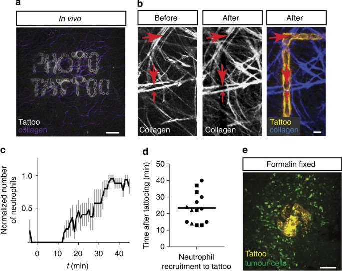
In vivo imaging and histochemistry are combined in the cryosection  labelling and intravital microscopy technique | Nature Communications
In vivo imaging and histochemistry are combined in the cryosection labelling and intravital microscopy technique | Nature Communications

Annexin-vivo | Application Support Knowledgebase | Lab Products & Services  | PerkinElmer
Annexin-vivo | Application Support Knowledgebase | Lab Products & Services | PerkinElmer

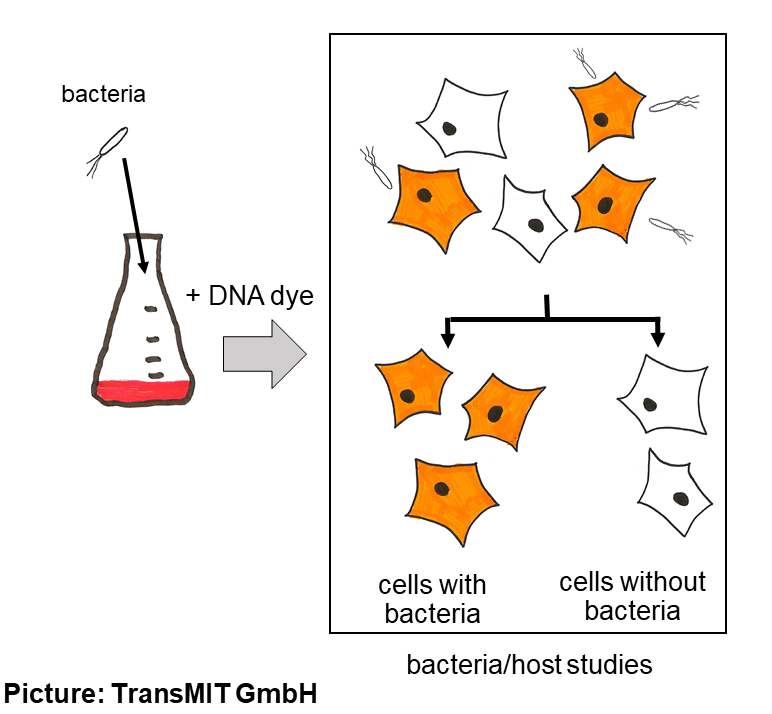
Cyanine dyes for in-vivo staining of microorganisms

Human Ex vivo Wound Model and Whole-Mount Staining Approach to Accurately  Evaluate Skin Repair | Protocol (Translated to Turkish)
Human Ex vivo Wound Model and Whole-Mount Staining Approach to Accurately Evaluate Skin Repair | Protocol (Translated to Turkish)

In vivo staining of cortical astrocytes with sulforhodamine 101... |  Download Scientific Diagram
In vivo staining of cortical astrocytes with sulforhodamine 101... | Download Scientific Diagram